

Supracondylar fracture occurs most
commonly in children between the age 5 and 15 years
It is caused by a fall on the out stretched
hand

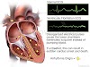
Basic anatomy :
The elbow joint is a simple hinge
joint allowing movement in only one plane
It is made up of three seperate
joints namely
- Proximal radioulnar joint
- Humeroradial joint
- Humeroulnar joint

The distal end of the humerus is flattened farming
two process namely the
- lateral epicondyle
- medial epicondyle

Medial to lateral condyle lies the
capitulum
Lateral to the medial condyle lies
the trochlea

The bony points
There are three bony points around
the elbow
- the medial condyle
- the lateral condyle
- the tip of olecranon

- They help in determining the normal anatomy of the joint.
- These reference points also helpful in determining any deformity of the elbow joint
- Normally in an elbow flexed at 90°, the three bony points make an isosceles triangle (any two sides are in equal length)
- In extended position of elbow, these three bony prominence make a straight line
The Carrying angle
When the elbow is extended and
supinated the long axis of the arm and that of forearm form an angle which is
called Carrying angle

- It disappears in flexion of elbow
- Normal angle
- Male : 11° Female : 14°
Ossification around the elbow
- One should be able to differentiate between the normal ossification centre and the fractured fragments

- without this knowledge one may think normal ossification centre as a fractured fragments
TYPES :
1.Extension type : 95-98%
In this type the distal fragments is
displaced backwards

2.Flexion type :
In this rare type the distal
fragments is displaced forward
CLINICAL FEAUTURES :

- The child complains of severe pain in the elbow and holds it in the flexed position
- The swelling is tense filling up the hollow around the elbow
- Palpitation will elicit tenderness in the distal end of the humerus

- In this injury one should always feel the radial pulse to see if there is any pressure in the brachial artery
- A weaker radial pulse compared to the opposite side needs emergency attention to save the circulation of forearm
DIFFERENTIAL DIAGNOSIS :
Posterior dislocation of elbow :
In supracondylar fracture the normal
triangle relationship is not disturbed in case of posterior dislocation the
relationship is grossly disturbed and the three points lies in a
line
DIAGNOSIS :
1.Physical exam :

- gross deformity
- swelling
- ecchymosis {bruises} in antecubital fossa
- limited elbow motion
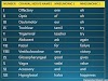
2.Nerve exam
evaluate for
Anterior interosseous nerve {ACN} neuroproxia
can't make OK sign

Median Nerve injury :
loss of sensation over the volvar
index finger

Radial nerve neuroproxia :
inability to extend wrist, MCP
joints,thumb joint

3.Vascular exam
- assess vascular perfusion
well perfused- warm and pink
- poorly perfused
cold and pale
4.Imaging
Recommanded views : AP
and lateral view of elbow
Findings :

- Positive
posterior fat pad sign
- Displacement
of the anterior humeral line
Capitulum moves posteriorly to this reference line in extension type and anterior in flexion type
- Alteration in Baumann angle
Baumann line is created by drawing a
line parallel to the longitudinal axis of the humeral shaft and a line along
the lateral condylar physis as viewed on AP

Normal 70-75°
but best judge by comparing it with the contralateral side
RADIOLOGICAL CLASSIFICATION :
Based on X ray, Garland has
classified supracondylar fracture as following
Type 1 : Undisplaced
Type 2 : Partially
displaced but posterior cortex intact
Type 3 : Complete
displacement in both anterior posterior and lateral views

TREATMENT :
1.Crack with displacement :
only a posterior plaster slab with
good padding for about 2-3 weeks

2.Displaced fracture :
- need reduction under general anaesthesia with traction , countertraction and local pressure
- a posterior slab is applied with good padding
3.If fracture fragments are unstable
:
open reduction and internal fixation
with K wire is done
4.Injury to the Neurovascular bundle :
A fracture complicated by any injury to the neurovascular bundle leading to
neurovascular compromise requires exploration and decompression with
appropriate vascular and plastic surgery inputs
COMPICATION :
Early :
- Injury to the median nerve {anterior interosseous nerve}
- Injury to the brachial artery {volkmann ischemia}
- Pin migration
- Infection
- Nerve injury
order of occurrence of nerve injury
:
1st Anterior interosseous nerve (branch of median
nerve )
2nd Median nerve –posterolateral displacement
3rd
Radial nerve - posteromedial
displacement
4th
ulnar nerve – commonly injured iatrogenically by pinning
Late :
1.Cubital varus deformity :

- Most common complication
- Malunion of the fracture leading to the cubitus varus deformity called Gunstock deformity
- Corrected by supracondylar osteotomy of humerus
2.myositis occificans

3.elbow stiffness

4.Cubital valgus (rare)

5.Volkmann ischemic contracture


0 Comments