

Definition:
COPD is a chronic pulmonary disease
characterized by persistent respiratory symptoms and airflow limitations which
is caused by small airway obstruction and parenchymal distruction.
Anatomy:

Epidimiology:
Male:Female ratio - 3:2
3rd most common cause of death worldwide
Etiology:
1. Environmental factors
Tabacco smoke -95%of case
Indoor airpollution
Occupational exposure. Eg: coal dust,silica
Lung malformations. Eg : childhood
infections,maternal smoking
Recurrent infection of lung
Low socioeconomic status
Cannabis smokking {inhalation of smoke by
heating the flowers,leaves or extract of cannabis}
2. Host
factors
Genetic factors – alpha 1-antitrypsi
deficiency
Airwayhyperreactivity
Types:
1.
Chronic
bronchitis
2.
Emphysema
1.CHRONIC BRONCHITIS: (blue bloaters)
Productive cough for at least 3months each year
for 2 consecutive years

2.EMPHYSEMA: (pink buffers)
Persistent dilation of pulmonary air space distal to the terminal bronchioles caused by distruction of the alveolar walls and the pulmonary capillaries required for gas exchange

Types of empysema:
1.Centriacinar emphysema:
Focal enlargement and destruction of
respiratory bronchioles
Alveoli unaffected
Seen in smokers,affect upper lobe
2.Paracinar (Panlobular) emphysema:
Destruction and enlargement of all portion of
the Acinus
Associated with alpha-1 antitrypsin
deficiencty
Affect lower lobe
3.Paraseptal (distal acinar) emphysema:
Enlargement and destruction of alveoli
Bronchioles are unaffected
4.Irregular (para-cicatrical) emphysema :
Associated with scaring
5.Mixed emphysema:
Consist of above two or more types

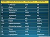
Difference between chronic bronchitis and emphysema
|

Pathophysiology:
1.Chronic bronchitis :

2.Empysema :

Clinical feautures :
Common
Chronic
cough with expectoration (sputum)
Dyspnea
and tachypnea
Pursed
lip breathing (to increase +pressure in lungs and prevent airway collapse)
End
expiratory wheezing ,crackles
Tachycardia
Cyanosis (bluish colouration on skin and mucous membrane)
Weight
loss and cachexia (weight loss and muscle wasting syndrome)
In
advanced COPD
Congested
neck veins (due to increased jugular venous pressure)
Barrel
chest
Asynchronous
movement of chest and abdominal during respiration
Use
of accessory respiratory muscles due to diaphragmatic dysfunction
Hyperresonant
lung
Decreased
breath sounds on auscultation (silent lung)
Prolonged
expiratory phase
Finger clubbing
1.Assessment :
age,history,clinical signs
2.Spirometery :
Spirometry is performed pre and post
bronchodilator admisteration to determine whether airflow limitation is
present,partial or fully reversible
COPD is confirmed when FEV/FVC ≤ 70%
Ø Pulseoxinometer –to detect O2 saturation
Dyspnea scale
|
RATIO |
CONDITION |
|
≥80% |
Mild |
|
50-79% |
Moderate |
|
30-49% |
Severe |
|
≤30% |
Very severe |
3.Blood gas
analysis BGA:
To find hypoxemia-absence of O2, in blood
Hypercapnia-increased CO2
in blood
Polycythemia-RBC elevated in
blood
4.Chest X-ray:
Shows signs of Barrel chest
[hyperinflated chest]
Decreased lung markings
Increase anterior ,posterior diameter
Diaphragm pushed down and flattened
Horizontal ribs and widened intercostal space

4.CT scan :
To evaluate complications
To rule out differential diagnosis
To plan surgery
5.Lab test :
Haemoglobin levels
Alpha-1 antitrypsin level
6.Gram strain :
In case of pulmonary
bacterial infection
7.Bronchoscopy :
procedure to look inside the lungs
8.ECG: shows signs of heart failure
Management :
1.Cessation of smoking
2.Bronchodilators : dilate bronchial walls
Shortacting
–for mild case Eg: Salmetrol,ipratropium
Long acting –for moderate to severe case
Eg: tiotropium bromide,formoterol
3.inhaled corticosteroids : lowers inflammation
Budesonide
Beclomethasone
Fluticasone
4.Vaccination :
Pneumococcal,influenza vaccination
5.Oxygen theraphy :
For patient with severe dyspnea
It increase the chance of survival in COPD
6.Pulmonary rehabilation :
Physiotheraphy
Pursed lip breathing,
Postural drinage
Physical activity to maintain
endurance,dyspnea
7.Vitamine D3 and calcium in case of
deficiency :
it reduce acute exerbration (worsening of symptoms)
8.Surgery :
Lung resection – Section of lung or entire lung is removed
For emphysema patients to improve
symptoms

Lung
transplant
For patients with non reparable lung damage
Complications:
1.Acute exerbation of Chronic obstructive
pulmonary disease (AECOPD)
Acute worsening of respiratory symptoms
Eg: icreased dyspnea,sputum consistency
changes
2.Chronic respiratory failure
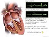
3.Corpulmonale (Right heart failure) :

4.Pulmonary cachexia (weight loss syndrome)
5.Secondary spontaneous pneumothorax –due to rupture of bullae (fluid filled sac)
Differential diagnosis :
Tuberculosis
Bronchiectasis
Asthma
Congestive heart failure
Lung cancer
 |
| Pink Puffers vs Blue Bloater |


1 Comments
Nice..
ReplyDeleteContinue......